We seek the big effect
Strategically focusing on rare diseases with defined genetic defects, BioMarin strives to develop innovative therapies that are first- or best-in-class.
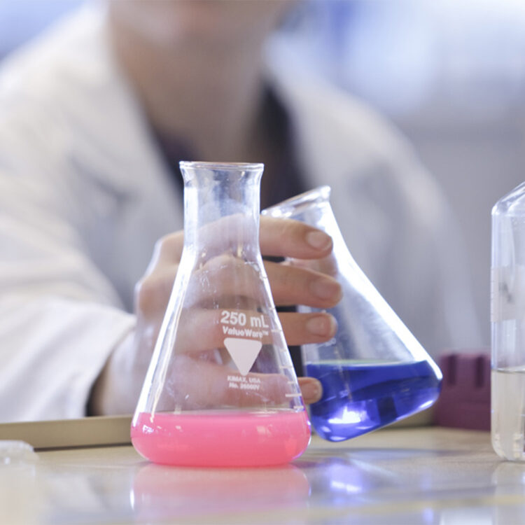
At BioMarin, we view ourselves as a team of pioneers, willing to take risks in the quest to find new solutions to address serious, debilitating challenges in rare diseases, and motivated by the desire to help improve the lives of patients with unmet medical needs.

BioMarin is committed to translating genetic discoveries into transformative medicines. Our approach focuses on diseases with precisely understood mechanisms that are rooted in GENETICS, developing TARGETED therapeutic interventions that address the underlying causes of diseases, utilising the readily ACCESSIBLE endpoints that are proximal to the disease pathway, and enabling TRANSFORMATIONAL changes to the way patients feel, function, and survive.
Please select a product to access further information including the Consumer Medicine Information and Product Information.
Registered pharmacies and hospitals only: Patients and consumers should speak to their community pharmacy or prescriber for more information.
DHL Direct to Market
Toll Free: 1800 077 421 (Option 1)
Email: d2mcs@dhl.com
Healthcare Logistics
Ph: 09 969 0734
E-mail: orders@healthcarelogistics.co.nz
Strategically focusing on rare diseases with defined genetic defects, BioMarin strives to develop innovative therapies that are first- or best-in-class.